Back Exercises for PIVD & Spinal Health
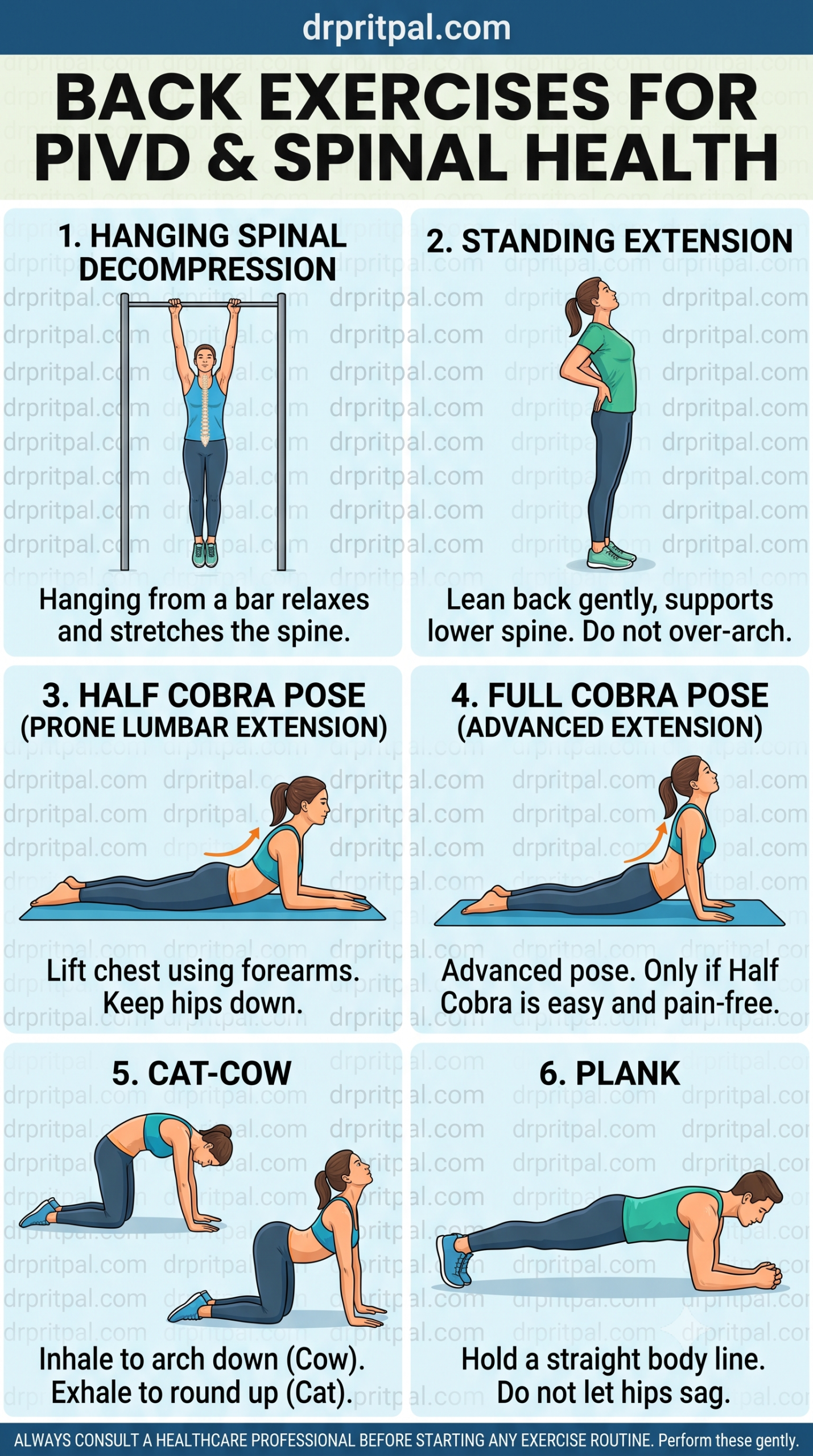
Reference this visual protocol overview detailing the 6 essential spinal decompression, extension, mobility, and core stabilization movements outlined below.
Acute Decompression & Nerve Root Relief
In the acute stage, the primary objective is to unload compressed lumbar intervertebral discs, minimize inflammatory chemical irritation on adjacent nerve roots, and avoid positions that worsen intradiscal pressure (such as sitting slumped or bending forward).
- PASSIVE SPINAL UNLOADING VIA GENTLE HANGING
- STANDING LUMBAR EXTENSION WITHIN COMFORT LIMITS
- STRICT AVOIDANCE OF FLEXION, TWISTING, AND PROLONGED SITTING
- SHORT-TERM NSAIDs OR ANALGESICS AS PRESCRIBED
1. Hanging Spinal Decompression
Using body weight to hang gently from an overhead chin-up bar helps create negative intradiscal pressure within the lumbar spine, widening neural foramina and relaxing paraspinal muscle spasms.
Gentle passive traction • 20 to 30 seconds per set2. Standing Extension
Standing upright with hands placed firmly on the posterior iliac crests for support, slowly lean backward into a gentle lumbar extension arc. This counteracts flexed postures and encourages anterior disc loading.
5 to 10 slow repetitions • Stop immediately if leg pain radiatesProne Extension & Centralisation Mobility
Grounded in Mechanical Diagnosis and Therapy (MDT) principles, repeated end-range extension movements assist in mobilizing the nucleus pulposus away from sensitive spinal nerves.
- HALF COBRA POSE (PRONE LUMBAR EXTENSION)
- CAT-COW SPINAL MOBILITY DRILLS
- MONITORING FOR PAIN CENTRALISATION PHENOMENON
3. Half Cobra Pose (Prone Lumbar Extension)
Lie face down on a flat exercise mat. Prop your upper body up on your forearms, keeping your pelvis and hips anchored firmly to the floor. Relax your lower back muscles and breathe smoothly.
Hold 15 to 30 seconds • Perform 3 to 5 repetitions5. Cat-Cow Mobility
Position yourself on all fours (hands under shoulders, knees under hips). Inhale deeply while gently dropping your stomach toward the floor (Cow), then exhale slowly while arching your spine upward toward the ceiling (Cat).
8 to 10 controlled cycles • Pain-free range onlyAdvanced Extension & Deep Core Stabilization
Once acute radicular symptoms are resolved and end-range extension is fully comfortable, rehabilitative focus shifts toward robust core muscular endurance to support spinal posture dynamically.
- FULL COBRA POSE (PROGRESSIVE END-RANGE EXTENSION)
- FOREARM PLANK (TRANSVERSE ABDOMINIS & LUMBO-PELVIC STABILITY)
- ERGONOMIC EDUCATION & LIFTING MECHANICS RETRAINING
4. Full Cobra Pose (Advanced Extension)
An advanced progression where you press up with fully extended arms, lifting your torso while maintaining complete pelvic contact with the floor. Proceed to this variation only if Half Cobra is entirely pain-free.
Progressive hold • Discontinue immediately if lumbar pinching occurs6. Plank (Core Stabilization)
Engage your deep abdominal core and gluteal muscles to hold a perfectly straight line from head to heels while supported on your forearms and toes. Prevent any downward sagging of the hips.
Hold 20 to 45 seconds • 3 progressive setsFrequently Asked Questions
What is the primary mechanical goal of PIVD rehabilitation?
The primary goal is to achieve pain centralisation—relieving distal radicular pain down the leg by shifting disc tissue away from compressed nerve roots using directional preference exercises.
Why is forward bending restricted during early recovery?
Lumbar flexion significantly increases pressure on the posterior intervertebral disc walls and can aggravate nerve root compression during acute disc prolapse phases.
How long does conservative recovery typically take?
Clinical studies show that the vast majority of symptomatic lumbar disc herniations resolve within 6 to 12 weeks with dedicated non-surgical management and structured rehabilitation.
When is surgical intervention required?
Surgery is considered when conservative measures fail after 2 to 3 months, or immediately if progressive motor weakness, neurological deficits, or cauda equina syndrome occur.
References
- Al Qaraghli, M. I., & De Jesus, O. (2023). Lumbar Disc Herniation. In StatPearls. StatPearls Publishing. https://www.ncbi.nlm.nih.gov/books/NBK560878/
- El Melhat, A. M., Youssef, A. S. A., Zebdawi, M. R., Hafez, M. A., Khalil, L. H., & Harrison, D. E. (2024). Non-Surgical Approaches to the Management of Lumbar Disc Herniation Associated with Radiculopathy: A Narrative Review. Journal of Clinical Medicine, 13(4), 974. https://doi.org/10.3390/jcm13040974
- Penchev, P., Ilyov, I. G., Todorov, T., Petrov, P.-P., & Traykov, P. (2024). Comprehensive Analysis of Treatment Approaches for Lumbar Disc Herniation: A Systematic Review. Cureus, 16(8), e67899. https://doi.org/10.7759/cureus.67899
- Sakai, D. (2022). The Essence of Clinical Practice Guidelines for Lumbar Disc Herniation, 2021: 4. Treatment. Spine Surgery and Related Research, 6(4), 329–332. https://doi.org/10.22603/ssrr.2022-0045
- Shinde, A. P. (2026). Evidence-Based Conservative Treatment Strategies for Lumbar Radiculopathy: A Systematic Review. PMC / Systematic Review, CRD420261407070. https://pmc.ncbi.nlm.nih.gov/articles/PMC13349735/